Sedimentology-Paleontology-Petroleum Exploration Facilities
• Sedimentology core laboratory
• Access to the Missouri Department Natural Resources McCracken core repository (located in Rolla)
• Rock preparation facility with assorted rock crushing, cutting, thin-section wafering, and polishing equipment, vacuum oven for porosity impregnation of epoxy resin into rock samples, and Frantz isodynamic magnetic separator
• Mineral Museum
• Paleontology Laboratory with extensive specimen and slide collection (>50,000) of catalogued invertebrate fossils, vertebrates, trace fossils, foraminifera, holothurians, and palynomorphs from various localities of different ages in the US, including the Gulf Coast, Mid-continent region, Western Interior Basin, Cordillerian region, Atlantic Coastal Plain, and around the world (e.g., West Africa, Germany, United Kingdom, South America, Australia)
• Microfossil sample preparation facility with vacuum and isolation hood and large-capacity and micro-centrifuges
• Hunt 3-D laboratory and remote sensing laboratory with more than 30 high-powered personal computers and software (including SMT Kingdom Suite) for teaching and research, in addition to Silicon Graphics MXE and SE Octane workstations with Landmark 3-D imaging system
• Modern Nikon, Leitz, Leica, Zeiss, and Vickers petrographic and reflected light microscopes, some with differential interference contrast
• Digital cameras and video recorder systems
• Zeiss Microscope Reflectance Photometer (MPMOIK)
• Research Devices infrared microscope
• Cathodoluminescence microscope system (Technosyn 8200 MkII and Nuclide ELM-2)
• Heating and freezing fluid inclusion stages (Fluid Inc. and Chaix Meca)
• Ultraviolet Fluorescence microscope
• Rigaku Miniflex x-ray diffractometer system with six-position sample interchange and Jade spectra software analysis system
• Perkin-Elmer Elan DRC-e Inductively Coupled Plasma/Mass Spectrometer (ICP/MS) with autosampler
• Perkin-Elmer Optima 2000 DV Inductively Coupled Plasma/Optical Emission Spectrometer (ICP/OES), ultrasonic nebulizer, dual view optics, and autosampler
• Newwave-Merchantek 213 nm laser ablation system for use with solid phase analysis with either the ICP/MS or ICP/OES systems
Facilities Within Other Units in the University
Facilities at the Graduate Center for Materials Research include:
• Philips EM430T Scanning Transmission Electron Microscope (STEM) equipped with back-scattered electron imagery and dispersive spectrometer
• JEOL T330A, Hitachi S-4700 FESEM, and Hitachi S570 scanning electron microscopes (SEM) equipped with back-scattered electron imagery and wavelength dispersive spectrometer
• Fully automated Scintag PAD V X-ray diffractometer and Scintag system computer processor and X-ray data library
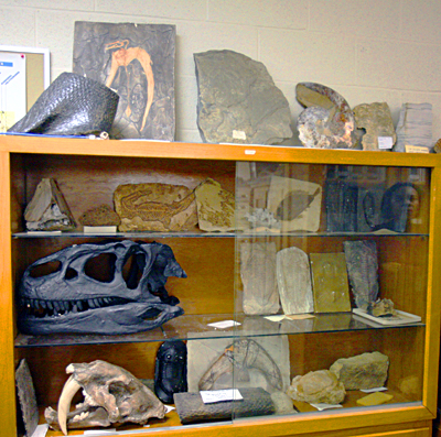
Part of the displays in the Paleontology Lab
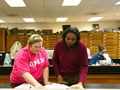
Professor Oboh-Ikuenobe (center) and her gradudate students in the Paleontology lab
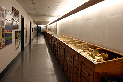
Collections in the Mineral Museum
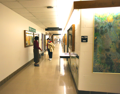
Professor Oboh-Ikuenobe talking with Marti Hanner (computer technicain) in the department hallway